- Home »
- Applications »
- Microscopy & Bioimaging »
- 2-Photon & Multiphoton Microscopy
2-Photon Microscopy
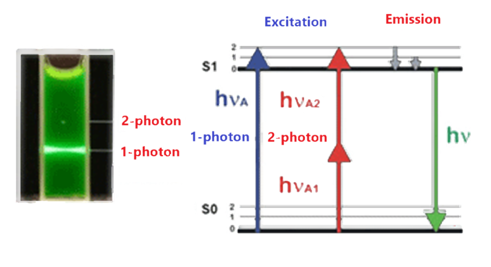
Nonlinear 2-photon excitation is based on the simultaneous absorption of two photons. Since the energy of a photon is inversely proportional to its wavelength, the two absorbed photons must have a wavelength about twice that of one-photon excitation. In 2-photon microscopy, two excitation photons from a pulsed laser (e.g., Ti:Sapphire or Nd:YLF laser) are combined to excite a fluorescent molecule. The molecule then emits a photon in the visible wavelength. 2-photon microscopy allows for out-of-focus background rejection similar to confocal microscopy.
The advantage of 2-photon microscopy over confocal microscopy is that it can penetrate deeper into tissue due to
- The absence of out-of-focus absorption
- The longer excitation wavelength and
- Less scattered light.
Nevertheless, the optical resolution is the same for both methods.
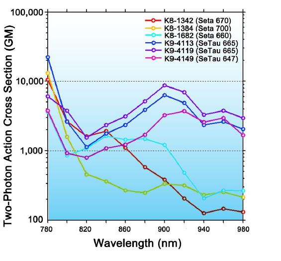
SETA BioMedicals has developed several green, red, and NIR emitting dyes (probes and labels) with very high 2-photon action cross sections (2PACS). Due to the donor-acceptor-donor structure of squaraine dyes, they exhibit much higher 2-photon absorption (2PA) efficiencies in comparison with other dyes [22-24] and, in particular, the new class of squaraine rotaxanes show extremely high 2PACS of up to 10,000 GM at near-infrared wavelengths critical for in vivo imaging (see below). Dyes and labels that undergo two-photon absorption (2PA) in the NIR and fluoresce in the far-red to NIR region with high quantum yields are very desirable to achieve deep tissue penetration.
and squaraine rotaxanes [24]
.jpg "\"2p-microscopy-neuron-BW(327x363).jpg\"")
While the 2PACS of common fluorophores are in the order of 10 - 200 GM (the 2PACS of the di-anioic form of fluorescein in water at pH 13 is 37 GM and for Rhodamine B the 2PACS is 204 GM for excitation at 830 nm in MeOH - see table below) and even some of the best 2P labels published have 2PACSs of only several hundred GM [23], Seta and SeTau dyes have 2PACSs in the order of thousand to several thousand GM in aqueous solution. SeTau and Seta labels are the best 2P probes and labels currently available on the market, combining high sensitivity and extremely high 2PACS: (K. Podgorski et al. Ultra-bright and -stable red and NIR squaraine fluorophores for in vivo two-photon imaging. PLoS One. 2012; 7(12): e51980).
The outstanding imaging properties of SeTau 647 are mentioned in the publication by the group of Prof. Weissleder at the Harvard Medical School: "Here we present the first generation of two-photon beta cell specific in vivo imaging probes based on GLP1R targeting peptides. Among the three compounds of potential interest, we found quite unexpectedly that a squaraine rotaxane conjugate (2PEx-647, K9-4148) had near-ideal in vivo imaging characteristics.
The relevant 2PA wavelengths for several Seta and SeTau dyes are provided in the table below.
Probes and Labels for in-vivo 2P-microscopy:
SETA BioMedicals has developed several 2P probes and labels with extremely high 2PACS and emission wavelengths between 500 and 700 nm. Seta-660, Seta-670, and, in particular, the squaraine rotaxanes (SeTau-665 and SeTau-647) are currently the most efficient 2P dyes available for the far red and NIR spectral emission range. SeTau-405, with good 2P excitation properties, is a dye with fluorescein emission characteristics. The 2P absorption wavelengths and cross-sections for these dyes are provided in the table below.
Seta and Squaraine rotaxane dyes offer unprecedented properties ideal for in vivo two-photon imaging. These dyes have two-photon cross-sections and photostabilities approaching those of quantum dots, but with molecular weights similar to other organic dyes, allowing labeling of subcellular structures, such as synapses, at low concentrations and with reduced functional impact. Importantly, they are non-toxic and allow long-term neuronal imaging. The demonstrated brightness and stability of these dyes promise to extend the limits of fluorophore concentration, imaging rate, illumination depth, and imaging duration for in vivo two-photon microscopy [24,42,43,44,45].
Below is a comparison of the 2PACSs for some popular dyes and our Seta and SeTau dyes:
| Product Number (Specs Sheet) |
Product Name (Product Info) |
Maximum 2PA Wavelength [nm] |
Emission Wavelength [nm] |
2P-Excitation Cross-Section [GM] |
|---|---|---|---|---|
|
-
|
Alexa 594
|
780 | 617 | 100 |
|
-
|
Alexa 647
|
1238 | 665 | 44 |
|
-
|
Alexa 700
|
1320 | 723 | 52 |
|
-
|
Cy5
|
1219 | 665 | 40 |
|
-
|
Fluorescein
|
770 - 790 | 515 | 37 |
|
-
|
Rhodamine B
|
830 | 620 | 204 |
|
-
|
Lucifer Yellow
|
840 | 528 | 1.3 |
|
-
|
Fura-2
|
700 | 512 | 11 |
|
-
|
Indo-1
|
700 | 475 | 1 |
| - |
DDTC
|
1550 | 785 | 105 |
| 750–850 | 518 | >100 | ||
| K8-1672 | Seta-646-NHS | 840 | 660 | 480 |
| K8-1384 | 800 | 705 | 1050 | |
| K8-1682 | Seta-660-NHS | 900 | 675 | 1625 |
| K8-1342 | Seta-670-NHS | 840 | 690 | 1925 |
| K9-4150 | SeTau-647 | 920 | 693 | >3000 |
| K9-4149 | 920 | 695 | 3700 | |
| K9-4119 | SeTau-665-NHS | 900 | 716 | >8500 |
2-Photon FRET:
2P-FRET microscopy has significant advantages over laser confocal FRET microscopy: while in confocal FRET microscopy, the actual FRET signal in the acceptor channel is always contaminated due to direct excitation of the acceptor molecule and the cross-talk between donor and acceptor emissions in the acceptor channel, in 2P-microscopy, it is possible to prevent the direct excitation of the acceptor molecule. 2P-microscopy also avoids out-of-focus bleaching, which allows repeated scanning in multiple focal planes and to obtaining a more detailed picture of intracellular events. 2P-FRET microscopy is also a powerful tool to assay intracellular protein-protein co-localization/interaction events.
Below is some information on the Forster radii, 2P-excitation wavelengths, and emission wavelengths of 2P-FRET pairs that are based on a combination of our most efficient 2P-donors with several of our acceptor dyes:
| 2P-Donor | Acceptor | Förster Radii (Å) |
2P-Excitation Wavelength [nm] |
Emission Wavelengths [nm] | |
|---|---|---|---|---|---|
| Donor | Acceptor | ||||
| 71–76* | 840 | 660 | 695 | ||
| 60–67* | 840 | 660 | 695 | ||
| 60–68* | 840 | 660 | 693 | ||
| 56–67* | 840 | 660 | 780 | ||
| 78–85* | 750–850 | 520 | 573 | ||
| 50–53* | 750–850 | 520 | 571 | ||
| 41–45* | 750–850 | 520 | – | ||
| 55–60* | 900 | 675 | – | ||
| 72–76* | 900 | 675 | 780 | ||
| 65 | 920 | 695 | - | ||
| 66 | 900 | 716 | - | ||
* Varies with Q.Y. of the donor